
Back to products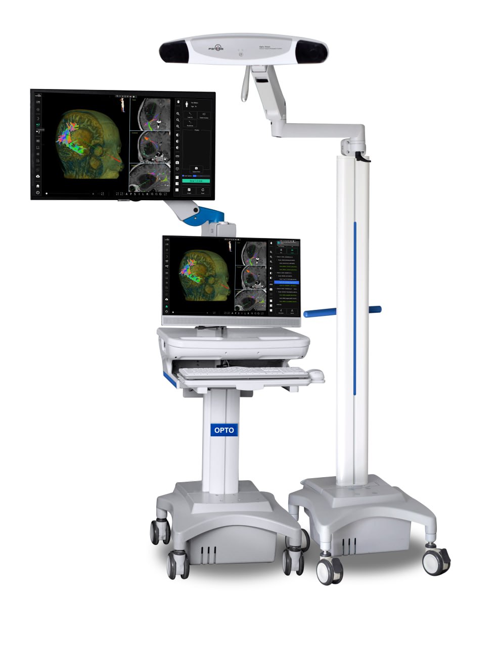
Products
OPTO
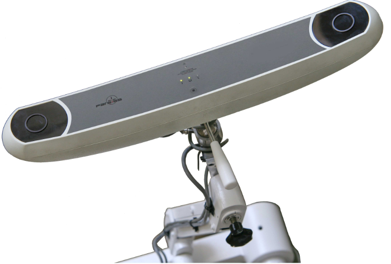
OPTO is a comprehensive, high-performance surgical navigation system for neurosurgery and ENT. Using advanced technology and a high-precision IR camera, it offers surgeons more accurate and reliable navigation during surgery.
Key Features
- Standalone planning station with full capabilities
- Compact design with high maneuverability
- Easy positioning of the surgeon's dedicated display
- Wide surgical coverage with long adjustable arms
Applications
Neurosurgery Applications
The brain is the most complex structure in the world, requiring precise surgical decisions and careful planning to minimize patient risk. The navigation system can be used for:
- Tumor biopsy
- Visualization of critical brain structures
- Tumor resection
ENT Applications
Like skull surgeries, ENT procedures require high precision to navigate very narrow pathways and cavities in this region. The navigation system in ENT can be used for:
- Sinus surgery
- Cyst and polyp removal
- Optic nerve decompression
Specifications
Image Data Transfer & Management
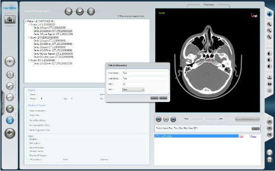
- Support for DICOM datasets including CT, MR, and PET
- Import from DVD, CD, and memory stick
- Review dataset tree structure before loading
- Preview image data before import
- Correct patient demographics recorded in DICOM
- Automatic detection of dataset issues
- Automatic compatibility check for navigation requirements
- Determine and correct medical image orientation from DICOM headers
- Add new datasets to a patient record intraoperatively
Visualization
- Easy imaging tools: zoom, rotate, and pan
- Window level, transparency, and color coding
- Full-screen image display
- Graphical patient orientation indicator
- Auto-hiding toolbars for maximum screen area
- Distance measurement
Image-to-Image Fusion
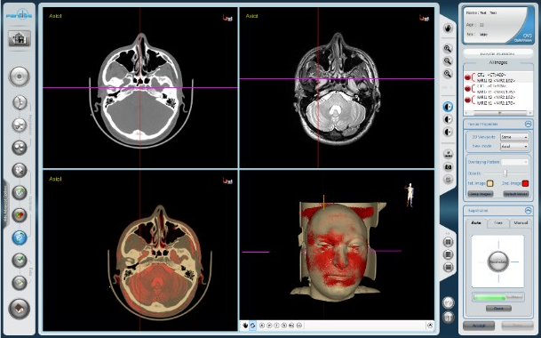
- Automatic fusion across SPECT, PET, CT, and MR (T1, T2, MRA)
- Manual refinement using anatomical landmarks
- Free manual adjustment by comparing two images
- Build volumetric models from fused images
- Adjust color and transparency of fused layers
- Combine fusion methods to achieve the best result
Segmentation & 3D Reconstruction
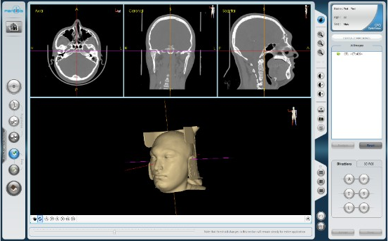
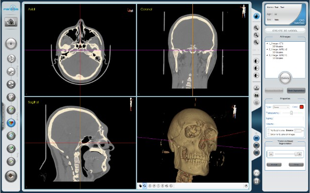
- Automatic bone and skin segmentation from CT
- Easy semi-automatic segmentation of selected regions
- Color-coded display of segmented structures
- Per-structure color and transparency control
- Specify VOI in 3D for segmentation
- Fast 3D reconstruction and standard cross-section views
- Define critical anatomical regions for surgical alerts
- Automatic volume calculation of segmented regions
Composite Modeling
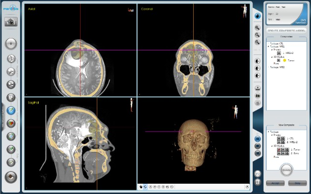
- Fast overlay of fused data and 3D models
- Individual opacity control per composite section
- Automatic validity check of composite models
- Automatic composite correction from updated fusion
- Create multiple composite models
Surgical Planning
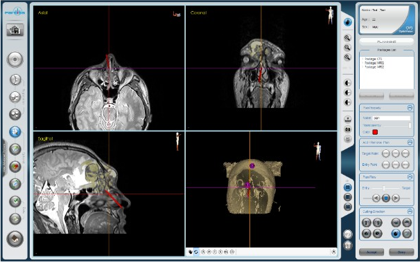
- Preoperative surgical simulation in a virtual environment
- Set and refine target and entry points on 2D or 3D models
- Adjust visualization parameters such as color and opacity
Tracking Camera & Tools
- Wide field of view
- Active and passive instrument support
- Automatic tool detection and validation
- Easy calibration and accuracy assessment
- Deactivate and reactivate individual tools
- Ergonomic instruments for surgical maneuvering
- Optimize tracker field of view with laser pointer
Registration
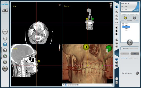
- Minimal-invasive registration workflow
- Automatic landmark suggestions by surgical region
- Manual landmark selection with 2D/3D refinement
- Display landmark error for registration refinement
- Automatic landmark capture
- Automatic evaluation of new landmarks
- Surface registration for higher accuracy
- Edit surface points to refine surface registration
- Report final registration error
Navigation
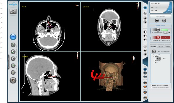
- Display tool orientation and tip in 2D and 3D
- Define check points for intraoperative registration verification
- Dynamic 3D model based on tooltip position
- Detect and compensate camera or patient displacement
- Concurrent endoscope and microscope video during navigation
- Freeze images for review, measurement, and other operations
- Display fused images in 2D and 3D
- Audio-visual alert when tracker loses reference or tools
- Alerts when approaching predefined critical regions
- Intraoperative simulation with virtual tool tip
- Real-time distance between targets and tooltip
- Auto-measure distance from tooltip to arbitrary points
- 3D distance measurement
- Review medical images during navigation
- Automatic full-screen mode
- Auto-snapshot of surgeon-defined views
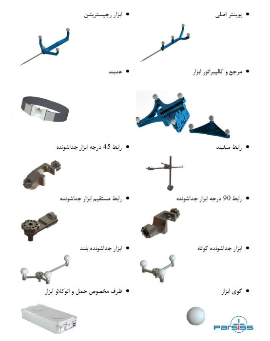
Accessories
- Main pointer, registration tool, and headband
- Tool reference and calibrator
- Mayfield adapter and 45°, 90°, and straight detachable adapters
- Short and long detachable tools and tool sphere
- Sterilization and transport case